Welcome to the Human Anatomy Lab Manual with Cat Dissections, a comprehensive guide designed for students to explore human anatomy through hands-on dissection experiences. This manual features 30 exercises covering all body systems, with clear instructions and full-color illustrations to enhance learning. Tailored for a one-semester course, it provides a structured approach to understanding anatomical concepts, ensuring a strong foundation in human anatomy. The manual aligns seamlessly with accompanying textbooks, offering a practical and engaging learning experience.
Importance of Anatomy in Medical Education
An understanding of human anatomy is foundational in medical education, providing the essential knowledge of how the body is structured and functions. Anatomy forms the basis for diagnosing and treating diseases, as it allows healthcare professionals to identify abnormalities and understand the relationships between different body systems. In the context of the Human Anatomy Lab Manual with Cat Dissections, hands-on dissection experiences bridge the gap between theoretical knowledge and practical application. By exploring anatomical structures, students develop critical thinking and observational skills, which are vital for future clinical practices. The study of anatomy also fosters an appreciation for the complexity and interdependence of body systems, enabling students to approach medical challenges with a holistic perspective. This lab manual emphasizes the importance of anatomy through detailed exercises, ensuring a robust foundation for aspiring healthcare professionals.
Overview of the Lab Manual Structure
The Human Anatomy Lab Manual with Cat Dissections is organized into a logical and comprehensive structure, designed to guide students through a detailed exploration of anatomy. The manual is divided into sections that cover external and internal anatomy, body systems, histology, and dissection techniques. Each chapter is supported by clear, full-color illustrations and step-by-step instructions, making complex concepts accessible. With 30 exercises, the manual ensures a thorough understanding of all body systems, from the skeletal and muscular systems to the nervous and circulatory systems. The lab manual is paired with customizable digital tools, such as flashcards and audio options, to enhance studying. Its engaging writing style and practical approach make it an invaluable resource for students, aligning seamlessly with accompanying textbooks for a cohesive learning experience.
Objectives of the Cat Dissection Course
The primary objective of the cat dissection course is to provide students with a hands-on understanding of mammalian anatomy, closely mirroring human anatomy. Through dissection, students develop essential skills in identifying and locating major organs and systems, fostering a deeper appreciation of anatomical relationships. The course aims to enhance observational and descriptive abilities, encouraging precise use of anatomical terminology. By exploring body systems such as the skeletal, muscular, nervous, and circulatory systems, students gain practical knowledge that reinforces theoretical concepts. Additionally, the course emphasizes safe dissection techniques and laboratory practices, promoting a respectful and professional approach to working with biological specimens. This experiential learning complements textbook studies, ensuring a well-rounded and comprehensive grasp of human anatomy principles. The ultimate goal is to prepare students for further studies in medicine, healthcare, and related fields with a solid anatomical foundation.
Preparing for the Anatomy Lab
Preparing for the anatomy lab involves gathering essential tools like scalpels, forceps, and gloves, and setting up a clean, well-organized workspace. Familiarize yourself with lab safety protocols, proper dissection techniques, and anatomical terminology to ensure effectiveness and efficiency during sessions. Organizing materials and reviewing the lab manual beforehand helps streamline the learning process, enabling a focused and productive experience. A prepared student contributes to a successful and safe laboratory environment, fostering academic growth and practical skill development. Proper preparation is key to maximizing the educational benefits of the anatomy lab.
Essential Lab Equipment and Tools
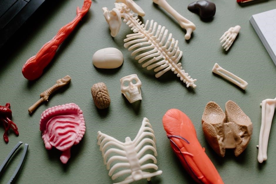
Essential lab equipment and tools are crucial for a successful anatomy lab experience. These include scalpels, forceps, dissecting scissors, and probes for precise dissection. Gloves, lab coats, and safety goggles protect students during procedures. Dissecting trays with wax or rubber lining help secure specimens, while magnifying lenses or microscopes aid in detailed observations. Additional tools like rulers, calipers, and anatomical charts enhance measurement and understanding. Proper equipment ensures safety, efficiency, and effective learning, allowing students to explore anatomy hands-on. Familiarizing oneself with these tools beforehand is vital for a smooth and productive lab session. The right equipment supports accurate dissection and fosters a deeper comprehension of anatomical structures, making it indispensable for the learning process in human anatomy.
Setting Up the Lab Environment
Setting up the lab environment is critical for a safe and effective anatomy lab experience. Begin by organizing workstations with essential tools and supplies, ensuring easy access. Dissection trays or pads should be placed centrally, with gloves, scalpels, and forceps arranged neatly. Proper ventilation is vital to minimize odors, and protective gear like lab coats and goggles should be worn. A clean, clutter-free workspace promotes focus and efficiency. Reference materials, such as anatomical charts or textbooks, should be within reach. Clearly label waste disposal areas for biological materials. Ensure all equipment is sterilized and ready for use. A well-organized lab environment fosters a productive and safe learning atmosphere, allowing students to focus on the dissection process and anatomical exploration. Instructor guidance is key to ensuring the setup meets safety and educational standards. Proper preparation enhances the overall lab experience and student engagement.
Identifying the Gender of the Cat for Dissection
Identifying the gender of the cat is essential for understanding its reproductive anatomy. Males can be identified by the presence of a scrotum, which contains the testes, and a prepuce, a small mound located anterior to the scrotum that houses the penis. Females have a vulva located posterior to the anus and possess mammary glands. Observing these external features aids in determining the cat’s gender. However, in some cases, the reproductive organs may have been removed, complicating gender identification. Using appropriate anatomical terminology and being aware of such variations during dissection is crucial. This step provides a foundation for accurately exploring the reproductive system and ensures a comprehensive understanding of feline anatomy.

External Anatomy of the Cat
The cat’s external anatomy includes fur, skin, and nails. Regional terms describe specific areas like the head, thorax, abdomen, limbs, and tail, aiding in precise anatomical descriptions and dissections.
Regional Anatomical Terms and Body Planes
Regional anatomical terms divide the cat’s body into distinct areas, such as the cranial (head), caudal (tail), ventral (abdomen), and dorsal (back) regions. These terms provide a standardized way to describe locations and structures during dissection. Body planes, including the sagittal (divides the body into left and right), frontal (or coronal, divides into front and back), and transverse (horizontal, divides into top and bottom), help in visualizing and describing the spatial relationships of organs and tissues; Understanding these terms and planes is essential for accurate communication and identification of structures during anatomy studies. They form the foundation for precise dissection and observation, ensuring consistency in learning and practice.
Understanding Directional Terms and Orientation
Directional terms are crucial for accurately describing the location and orientation of structures during cat dissection. Terms like proximal (closer to the origin) and distal (farther from the origin) help describe positions relative to a starting point. Anterior refers to structures toward the front, while posterior denotes those toward the back. Dorsal (toward the back) and ventral (toward the belly) describe positions relative to the body’s surface. Understanding these terms allows precise communication and ensures clarity when identifying anatomical features. Proper orientation is equally important, as it provides a consistent reference frame for dissection. Mastery of directional terminology and spatial awareness enhances the learning experience, enabling students to navigate the complexities of feline anatomy effectively and apply these principles to human anatomy studies. This foundation is vital for accurate and efficient dissection practices.
Internal Anatomy of the Cat
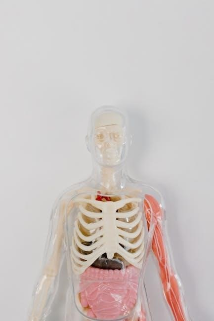
The internal anatomy of the cat reveals intricate body cavities and organs, providing insights into mammalian systems. Dissection exposes major organs like the heart, lungs, and digestive structures, aiding comparative studies with human anatomy.
Let me know if you’d like me to continue with another section!
Body Cavities and Membranes
Understanding body cavities and membranes is crucial for exploring the internal anatomy of the cat. The thoracic cavity contains the heart and lungs, while the abdominal cavity houses digestive organs. These cavities are lined by membranes such as the pleura and peritoneum, which reduce friction and protect organs. The diaphragm separates the thoracic and abdominal cavities, playing a key role in respiration. Students learn to identify these structures during dissection, gaining insights into their functions and relationships with major organs. This knowledge is vital for comparative studies with human anatomy, as similar cavities and membranes are present in humans. Proper identification of these features is essential for a thorough understanding of both feline and human internal anatomy.
Identifying Major Organs and Their Locations
In the cat dissection, identifying major organs and their locations is a critical step in understanding internal anatomy. The heart, located in the thoracic cavity, pumps blood through the circulatory system. The lungs, situated on either side of the heart, are essential for respiration. The liver, the largest organ, resides in the abdominal cavity and plays a key role in metabolism and detoxification. The stomach, small intestine, and large intestine comprise the digestive system, with the pancreas and gallbladder aiding in digestion. The kidneys, located near the spine, filter waste from the blood, and the spleen, near the stomach, aids in immune function. Accurately identifying these organs and their positions helps students understand their functions and relationships, providing a foundational knowledge applicable to both feline and human anatomy.

Body Systems Exploration
Exploring body systems through cat dissection provides a hands-on understanding of their structure and function. This includes the skeletal, muscular, nervous, circulatory, respiratory, digestive, urinary, and reproductive systems, offering insights into their interconnected roles in maintaining life and overall physiology.
Skeletal System: Bones and Joints
The skeletal system exam involves identifying bones and joints in the cat. Students will locate and name major bones, such as the femur, humerus, and vertebrae, and observe their structures. Joints, like the shoulder and knee, will be dissected to understand their anatomy. This exercise provides a detailed understanding of how bones and joints function in movement and support, mirroring human anatomy. The manual includes full-color illustrations to aid in identification, making complex structures easier to comprehend. By exploring the skeletal system, students gain foundational knowledge essential for advanced studies in anatomy and related fields.

Muscular System: Major Muscle Groups
This section focuses on the identification and exploration of major muscle groups in the cat, providing a comparative basis for understanding human musculature. Through dissection, students will examine the structure and arrangement of muscles, such as the flexors, extensors, and muscles of the limbs. The lab manual includes detailed illustrations and step-by-step instructions to facilitate the identification of each muscle group. By observing the attachments and actions of these muscles, students will gain insights into their functional roles in movement and posture. The exercises emphasize the relationship between muscles and bones, highlighting how the muscular system interacts with the skeletal system to enable locomotion and other bodily functions. This hands-on approach reinforces anatomical concepts and enhances the understanding of muscle anatomy in a practical and engaging manner.
Nervous System: Brain, Spinal Cord, and Nerves
This section delves into the exploration of the feline nervous system, offering insights into its structure and function. Students will dissect and examine the brain, spinal cord, and peripheral nerves, gaining a deeper understanding of their roles in controlling bodily functions. The lab manual provides detailed diagrams and descriptions to aid in identifying key nervous system components. By comparing the cat’s nervous system to the human nervous system, students can better comprehend the universal principles of neural anatomy. Hands-on dissection allows for the observation of nerve pathways and their connections to muscles and organs, highlighting the nervous system’s role in coordination and control. This practical approach enhances the learning experience, making complex anatomical concepts more accessible and engaging for students. The exercises are designed to promote a thorough understanding of the nervous system’s intricate structures and their importance in overall physiological function.
Circulatory System: Heart and Blood Vessels
This section focuses on the circulatory system, emphasizing the heart, blood vessels, and their roles in maintaining life. Through dissection, students will examine the feline heart, identifying its chambers and valves, and explore the structure of arteries, veins, and capillaries. Comparisons with human anatomy highlight similarities and differences, aiding in understanding blood circulation principles. The lab manual provides detailed illustrations and step-by-step guides for locating major blood vessels, such as the aorta, pulmonary arteries, and veins. Hands-on activities allow students to trace blood flow through the heart and vessels, demonstrating the system’s function in oxygen delivery and nutrient distribution. This practical approach, supported by clear instructions and visual aids, helps students grasp the circulatory system’s complexities and its essential role in overall physiology. The exercises are designed to foster a comprehensive appreciation of cardiovascular anatomy and its significance in maintaining bodily functions.
Respiratory System: Lungs and Airway Structures
This section delves into the respiratory system, focusing on the lungs and airway structures in the cat model. Students will dissect and examine the trachea, bronchi, and lungs to understand their roles in gas exchange. The lab manual provides detailed illustrations and instructions for identifying the larynx, tracheal rings, and bronchioles. Through hands-on exploration, students will learn how air moves through the respiratory tract and how oxygen is transferred into the bloodstream. Comparisons with human anatomy highlight similarities, such as the structure of alveoli and the function of the diaphragm in breathing. Practical exercises include tracing the pathway of air and analyzing the histology of lung tissue. This section aims to enhance understanding of the respiratory system’s structure and function, preparing students for further study in human anatomy and physiology. The exercises emphasize the importance of the respiratory system in maintaining life and overall health.
Digestive System: Organs and Their Functions
This section explores the digestive system, focusing on the organs and their roles in breaking down food into nutrients. Through cat dissection, students will examine the oral cavity, esophagus, stomach, small intestine, and large intestine. The lab manual provides detailed instructions for identifying the liver, pancreas, and gallbladder, highlighting their functions in digestion. Students will learn how enzymes and acids process food and absorb nutrients. Comparisons with human anatomy emphasize similarities, such as the structure of the stomach lining and the role of the liver in detoxification. Practical exercises include tracing the digestive pathway and analyzing tissue samples. This section aims to deepen understanding of the digestive system’s complexity and its vital role in nutrient absorption and waste elimination. The hands-on approach helps students connect anatomical structures with physiological processes, enhancing their grasp of human anatomy and function.
Urinary System: Kidneys and Urinary Bladder

This section focuses on the urinary system, with an emphasis on the kidneys and urinary bladder. Through cat dissection, students will explore the structure and function of these organs. The kidneys, located in the retroperitoneal space, are responsible for filtering blood, removing waste, and producing urine. The urinary bladder, a hollow muscular organ, stores urine until it is expelled. The lab manual provides step-by-step instructions for identifying the kidneys, ureters, and bladder, as well as their connections. Students will learn about the renal cortex and medulla, nephrons, and the role of the urethra in urine expulsion. Comparisons with human anatomy highlight similarities in kidney function and bladder structure. Practical exercises include dissecting the kidneys to observe their internal anatomy and understanding the process of urination. This section enhances students’ understanding of the urinary system’s role in maintaining homeostasis and overall health.
Reproductive System: Male and Female Anatomy
This section delves into the anatomy of the reproductive systems in cats, comparing both male and female structures. The male reproductive system includes the testes, epididymis, vas deferens, and penis, while the female system comprises the ovaries, oviducts, uterus, and vagina. Through dissection, students will identify these organs and their relationships. The lab manual highlights the role of each structure in reproduction, such as sperm production in males and egg development in females. Comparisons with human anatomy emphasize functional similarities, such as the role of the uterus in supporting embryonic development. Practical exercises include locating the reproductive organs, understanding their blood supply, and examining histological samples of reproductive tissues. This section provides a comprehensive understanding of the reproductive system’s structure and function in cats, aligning with human anatomy for a comparative learning experience.

Histology and Microscopic Anatomy
This section introduces histology and microscopic anatomy, focusing on tissue structure and cellular details. Students learn to prepare specimens for microscopy and analyze tissue types, linking macroscopic anatomy with microscopic observations.
Preparing Tissue Samples for Microscopic Examination
Preparing tissue samples for microscopic examination is a critical step in histology. Fixation preserves tissues, maintaining their structure and preventing decay. Sectioning involves slicing fixed tissues thinly for slides. Staining enhances visibility under a microscope, differentiating cell types and structures. Digital tools in eTextbooks, such as flashcards and highlighting features, aid in understanding these processes. This section guides students through proper techniques, ensuring accurate microscopic analysis and linking macroscopic anatomy to cellular-level observations. Clear instructions and full-color illustrations support hands-on learning, aligning with exercises covering all body systems. This foundational skill is essential for exploring human anatomy in depth.
Identifying Basic Tissue Types Under the Microscope
Identifying basic tissue types under the microscope is fundamental to understanding histology. The four primary tissues—epithelial, connective, muscle, and nervous—each exhibit distinct structural features. Epithelial tissues form layers, often lining surfaces, while connective tissues provide support and include components like collagen and blood vessels. Muscle tissues are characterized by their elongated cells and contractile properties. Nervous tissues contain neurons and supportive cells, facilitating communication. Proper staining techniques enhance visibility, allowing students to differentiate these tissues. This section provides detailed descriptions and high-quality images, enabling students to recognize and classify tissues accurately. By mastering this skill, students build a solid foundation for understanding the microscopic structure of human anatomy, complementing their macroscopic dissection studies. This exercise is crucial for correlating tissue structure with function in various body systems.
Dissection Techniques and Safety
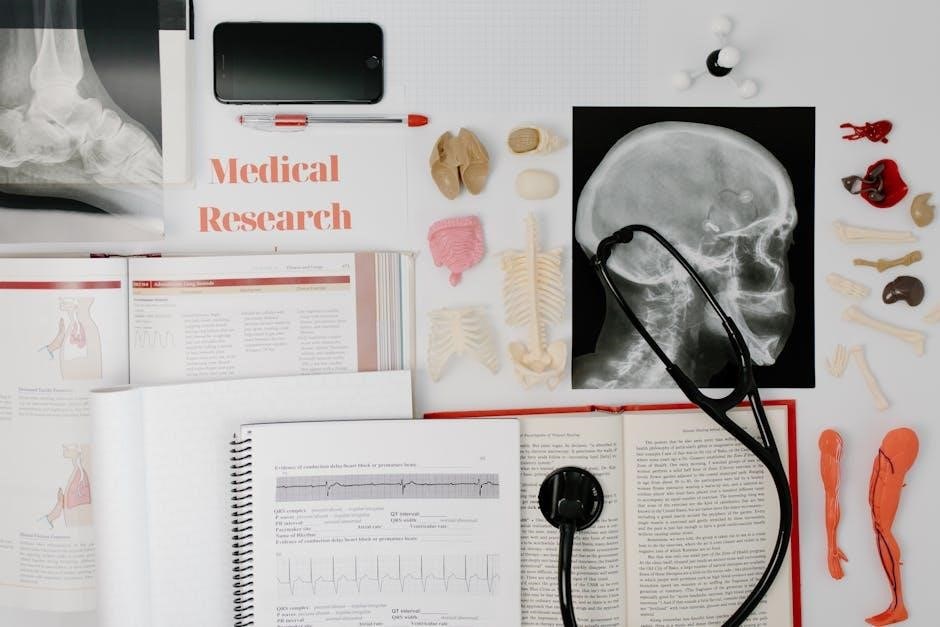
Dissection techniques involve precise proper use of scalpels and forceps. Safety requires protective gear and a clean workspace. Following protocols prevents accidents, ensuring effective dissection.
Step-by-Step Dissection Guide for Cat Anatomy
Begin by preparing the cat specimen on a dissecting tray, ensuring proper ventilation and safety gear. Start with an external examination to identify key landmarks. Make precise incisions to expose superficial muscles, carefully avoiding damage to underlying structures. Proceed to internal organs, systematically exploring body cavities such as the thoracic and abdominal regions; Use forceps and scalpels to locate and identify major organs like the heart, lungs, liver, and kidneys. Document observations and label structures for reference. Follow detailed instructions to minimize tissue damage and maintain anatomical integrity. Ensure all dissection steps align with anatomical terminology and laboratory protocols. This guide provides a structured approach to understanding feline anatomy, mirroring human anatomical principles. Adhere to safety guidelines throughout the process to ensure a successful and educational dissection experience.